СІИё: ШЛѓ -- РќЙЎАЁПы
УЪБт ШЛѓФЁЗсРЧ СпПфМК Йз УжМвРЧ ШЛѓШфХЭИІ АЁСЎПРДТ ШЛѓ УЂЛѓРЧ УжБйРЧ ФЁЗс[ Early
Burn Wound Management] : Revised July 1st 2007
Dong-Chul, Kim M.D., Ph.D
ШЛѓРЛ РдРКШФ ШЛѓШфХЭИІ ГВБтСі ОЪЕЕЗЯ ЧЯБтРЇЧиМДТ УЪБт ШЛѓРЧ ФЁЗсАЁ ИХПь СпПфЧбАЭРЬ АСЖЕЧАэ РжРИИч, АЁДЩЧб ЛЁИЎ УЪБтШЛѓУЂЛѓРЛ
ФЁРЏ НУХААХГЊ ШЛѓКЮРЇИІ СйРЬДТ АЭРЬ АсСЄРћРЬДй.
УЪБт ШЛѓ ШЏКЮ ЕхЗЙНЬПЁМ АЈПА Йз ПАСѕРЧ СЖР§, НРРБ ШЏАцРЧ РЏСі, ЧЧКЮ РчЛ§РЛ ЕНДТ МКРхРЮРк, ЛчРЬХфФЋРЮРЧ ХѕПЉ, БЙМвРћ Heparin ФЁЗсРЧ НУРлЕюРЬ АСЖЕЧАэ РжДй.
РЬЗЏЧб ФЁЗсРЧ ModalityИІ РЇЧиМ УжБй АЈПАСЖР§РЛ РЇЧиМ ПРПАЕЧАэ ПАСѕ ЙнРРРЬ РжДТ ШЏКЮПЁМ with ActicoatРЧ ЛчПы, 2ЕЕ ШЛѓЕюРЧ НЩКЮШЛѓРИЗЮРЧ РќШЏ[conversion]РЛ ЙцСіЧЯДТ БЙМвРћl heparinРЧ ЛчПы, - antithrombotic and antiinflammatory effects , effects of increased extracellular glycosaminoglycans which enhance collagen synthesis and epithelializationЕюРЬ РжРН], РЬПмПЁ ЧЧКЮРЧ КќИЅ РчЛ§ПЁ ПЕЧтРЛЙЬФЁДТ МКРхРЮРк ЕщРЧ ЛчПы[EGF, PDF,placental extracts(LaennecЈо)ЕюРЛ ШЛѓ РќЙЎАЁРЧ ОіАнЧб УЂЛѓАќИЎЧЯПЁ ЛчПыЧвМі РжДй.
УжБйРЧ РЬЗЏЧб АГГфРЧ РгЛѓ МКАј ЛчЗЪАЁ КИАэЕЧАэ РжРИИч, РЬДТ АэНФРћ ФЁЗсПЁ КёЧЯПЉ КЮКаУў ШЛѓШЏРкРЧ УЂЛѓФЁРЏ БтРЯРЬ 1-3РЯ СЄЕЕ СйОюЕхДТ АсАњИІ КИРЬАэ РжОю ЕћЖѓМ ЙпЛ§ЕЧДТ ШЛѓШфХЭИІУжМвШ Чв Мі РжДйДТ СЁРЛ СіРћЧвМі РжДй.Ч
Indication: ЛчПыРЧ РћРРСЖАЧ
1) contaminated or infected burn wound over mid second degree to thisr degree burn, the ActicoatTM was used from the acute stage.
2) In the cases which usually burn wound showed excoriation of bullae and relatively non infected wound, the topical heparin or placental extracts therapy were started. For using topical heparin therapy, the heparin solution, 100-200U/ml was prepared, and it moisten the the hydrophilic moist wound healing products such as AquaCelЈо or MediformЈоTM ]. After these dressing materials soaked in the heparin solution were applied on the denuded burn wound, and it overdressed with transparent film dressing such as TegadermЈо or Op siteЈо dressing for keeping the moist wound environment.
3) Also In the cases which usually burn wound showed excoriation of bullae and relatively non infected wound, one of growth promoting agent of epithelialization, placental extract [LaennecЈо ]
Fo rits use, after 2 ml of LaennecЈо was diluted in 20
ml saline, it can be made 10% LaennecЈо solution. It moisten the hydrophilic
moist wound healing products for containing of placental extracts. The
soaked moist wound products were applied over the burn wound, and it also
covered with transparnet film dressing for keeping the moist wound environment.
АЂЗа =
П МеЛѓ (THERMAL INJURIES)
ПТЕЕПЁ РЧЧб ЧЧКЮРЧ ЦФБЋДТ НЩАЂЧб БЙМвРћ БзИЎАэ РќНХРћ КЏШИІ УЪЗЁЧбДй. РЬЗЏЧб ЧЧКЮСЖСї ЦФБЋДТ П, ШЧа ЙнРР, РќБт, ШЄРК ЧбЗЉПЁ РЧЧи РЯОюГДй. НЩЧб П МеЛѓРЛ РдРК ШЏРкРЧ УГФЁПЁДТ БЙМвРћ ЧЧКЮМеЛѓРЧ КДХТЛ§ИЎЧа, СјДм, УГФЁЛг ОЦДЯЖѓ ПМеЛѓРИЗЮ РЮЧб РќНХРћРЮ ЧїОз ПЊЧаРћ(hemodynamic), ДыЛчРћ(metabolic), ПЕОчЧаРћ(nutritional), ИщПЊЧаРћ(immunologic), БзИЎАэ СЄНХРћ ЧзЛѓМК(psychological homeostasis) БтРќЕюРЧ КвБеЧќПЁ ДыЧб РЬЧиАЁ ЧЪПфЧЯДй.
БЙГЛ ШЛѓШЏРкРЧ ЙпЛ§РК ИХГт Ор 13000Иэ РЬЛѓРЬ РдПј АЁЗсЧЯАэ РжРИИч, Ор 23ИИИэРЬ ПмЗЁШЏРкЗЮ КДРЧПјПЁМ ФЁЗсЕЧАэ РжДй. БЙГЛ РдПјЧб ШЛѓРЧ АЁРх ИЙРК ПјРЮРИЗЮДТ ШПАШЛѓРЬИч, ПХСШЛѓ, СЂУЫШЛѓ, РќБтШЛѓ, ШЧаШЛѓ БтХИРЧ МјРЬДй. МвОЦШЛѓШЏРкДТ РќУМШЛѓШЏРкРЧ Ор 1/3РЛ ТїСіЧЯАэ РжДй. УжБй 6АГПљПЁМ 2Гт ЛчРЬРЧ ПЌЗЩРЧ МвОЦПЁМ РќБт ЖЧДТ ОаЗТ МмРЧ СѕБтПЁ РЧЧи МіКЮПЁ ШЛѓРЛ СѕБтШЛѓРЧ ПЙАЁ БЙГЛПЁ ИЙРЬ ЙпЛ§ЕЧАэ РжОю КЮИ№РЧ АЂКАЧб СжРЧАЁ ПфБИЕШДй. ЖЧЧб 2 - 6ММРЧ ОюИАОЦРЬПЁМ ГУПТМіБтПЁ РЧЧб ШЛѓ ЖЧЧб КѓЙјЧЯАд РЯОюГЊАэ РжДй.
ШчШї ШЛѓШЏРкРЧ РдПјБтАЃРК РќНХУМЧЅИщРћ(total body surface area: TBSA)ПЁ ДыЧб ШЛѓКЮРЇРЧ %ПЁ 1-1.5РЯРЛ АіЧб БтАЃРЬ ПфЧбДй.
ЁЁ
I. ШЛѓ(BURNS)
ЁЁ
1) ЧЧКЮРЧ БтДЩ Йз БИСЖ(function and structures)
(1)БтДЩ
ЙцОю(protection): СжКЏ ШЏАцРЧ ПТЕЕ, ШЧаЙАСњ, ММБе, ПмЛѓ ЕюПЁ ДыЧб КИШЃКЎ
ИщПЊЧаРћ(immunologic): АЂСњУў(keratin layer)РК ММБеПЁ ДыЧб ЙцОю РлПыРЛ ЧбДй.
МіОз БеЧќ(fluid balance): МіКа МвНЧ(water loss)РЛ ЙцСі
УМПТСЖР§(thermoregulation): АњЕЕЧб П МвНЧАњ П ШЙЕцРЛ ЙцСі
НХАцАЈАЂ(neurosensory); ХыСѕ,УМПТ, АЈАЂРЧ МіПыБт(receptors)
ЛчШИРћРЮ ЛѓШЃРлПы(social-interaction): ПмИ№
(2)БИСЖ
a)ЕЮВВ: Ор 1-2 mm РЬИч, ЙЬЙпДоЕШ РЏОЦ, РЇУрРЬ РжДТ ГыРЮПЁМ ОуОЦСјДй.
b)ЛѓЧЧ: ЙйБљТЪ ОуРК УўРИЗЮ СжЗЮ ЛѓЧЧММЦїЗЮ БИМКЕЧОю РжРИИч, СІРЯ
ЙйБљТЪПЁ ФЩЖѓЦО(keratin)РЛ ЦїЧдЧб ММЦїИІ АЂШММЦї(keratinocyte)Жѓ КЮИЅДй.
БтРњ ЛѓЧЧММЦїДТ ЦФРЬКъИЎГыГиЦО(fibronectin)РЬЖѓ КвИЎДТ КЮТј КаРк(adhesion molecule)ПЁ РЧЧи БтРњИЗ(basement membrane)ПЁ КЮТјЕЧОю РжДй. РЬ ЙЬМКМї ММЦїДТ АшМг КаПЧЯПЉ РЬЕП(migration) ЕЧОю ЙйБљТЪРИЗЮ ХЛАЂЕШ ЧЅИщРЧ ММЦїИІ КЙРЇЧбДй. ПмЛѓШФ ЛѓЧЧММЦїРЧ КЙРЇ(replacement)ДТ РЬЗЏЧб РчЛ§АњСЄ(regenerative process)ПЁ РЧЧЯИч, РЬЗЏЧб РчЛ§РК УжРћРЧ СЖСї ФЁРЏ ШЏАц(optimal tissue healing enviroment)ПЁ РЧЧиСіИч, МКРхРЮРк(growth factor)ЖѓАэ КвИЎПьДТ ШЧаРћ РкБи(chemical stimuli)РК ЙАЗа ММЦї КЙСІ(replication), РЬЕП(migration)РЧ АњСЄРИЗЮ РЬЗчОюСјДй. ИЙРК Бз НХШЃ(cues)ДТ СјЧЧ ПфМв(dermal elements)ПЁМ ПРИч ЦЏШї СјЧЧРЧ БтСњ ДмЙщСњ(matrix protein)РЮ ЦФРЬКъИЎГыГиЦО(fibronectin), БтСњ ШЧеЙА(matrix compound)РЮ hyaluronic acidЗЮКЮХЭ ПТДй.
(c)СјЧЧ: РЏЕЮ СјЧЧ(papillary dermis) -- epidermal rete pegРЛ ЦїЧд
ИСЛѓ СјЧЧ(reticular dermis)
БтКЛ ММЦї Чќ(primary cell type)РК МЖРЏОЦММЦї(fibroblast)РЬИч, ПЉБтПЁМ СпПфЧб ММЦїПм БтСњ ДмЙщСњ(extracellular matrix protein)РЮ БГПјСњ(collagen), ХКЗТМв(elastin), БтСњ(matrix,ground substance)ЕюРЛ Л§МКЧбДй. ЖЧЧб РЬ ММЦїДТ ЛѓЧЧММЦїИІ БтРњММЦїИЗПЁ КЮТјНУХАДТ КЮТјДмЙщСњ(adhesion protein)РЛ Л§ЛъЧбДй. РЬ КЮТј ДмЙщСњ(adhesion protein)РК ЛѓЧЧРЬЕП(epidermal migration)Ањ КЙСІ(replication)ПЁ РлПыЧЯБтЕЕ ЧбДй. БтСњ(matrix)РК glycosaminoglycans(GAG component) ЖѓАэ КвИЎДТ complex polysaccharide-protein complex, hyaluronic acidЕюРИЗЮ РЬЗчОюСЎ РжДй. РЬ БтСњ(matrix)РК ММЦї(cell)ПЭ АсУМСЖСї ЙцЧтМК( connective tissue orientation) ММЦїПЁ ПЕОчКа ШЎЛъ(nutrient diffusion)РЛ ЧЯАдЧЯДТ ЙнРЏЕПМК(semifluid)РЛ СІАјЧЯАэ, ММЦї РЬЕП(cell migration)РЧ КёАш(scaffold)ЗЮ ПЊЧвРЛ ЧбДй. МЖРЏОЦММЦїПЁМ Л§ЛъЧЯДТ ЙАСњ(Fibroblast products)ЗЮДТ Јч БГПјСњ(collagen, СжЗЮ ЧЧКЮПЁМДТ Type one) Јш БтСњ ДмЙщСњ(matrix protein)ЗЮДТ ЦФРЬКъЗЮГиЦО(fibronectin), tenascin Ею Јщproteoglycans, glycosaminoglycan, hyaluronic acid, БтХИРЧ ДйИЅ БтСњ БИМКМККаЕщ(matrix components), ЈъЛчРЬХфФЋРЮ(cytokynes), ДйИЅ МКРх УЫСјЙАСњ( other growth stimulants)ЕюРЬ РжДй.
ЁЁ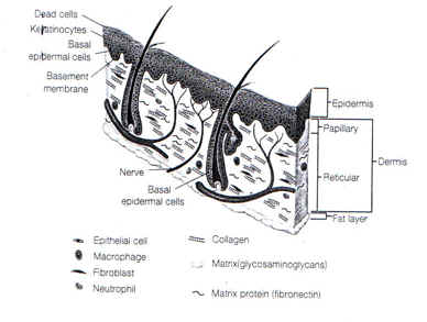
Fig.1. ЧЧКЮРЧ БИСЖ
ЁЁ
2) ШЛѓШФ Л§ИЎЧаРћ КЏШ
a)МјШЏБт КЏШ(circulatory derangement)
И№ММЧїАќ ХѕАњМК(capillary permeabilty)РЬ СѕАЁЧЯИч, ДмЙщСњ(protein)Ањ РќЧиСњ(ЦЏШї Na+) РЬ ИЙРК УМОз МеНЧРЬ ЙпЛ§ЕЧИч ЧїОзЗЎАЈМв(hypovolemia), НХЧїЗљЗЎ(renal blood flow) АЈМв, GFR АЈМв, КѓДЂ(oliguria), venous return , НЩЙкУтЗЎ(cardiac output) АЈМв, ЧїОзЗЎ АЈМвПЁ РЧЧб Мя(hypovolemic shock)ЕюРИЗЮ СјРќЕШДй.
b)КѓЧї(anemia)
ЦђБе РћЧїБИ МвНЧРЬ РќУМРЧ 15%
(РќУМ РћЧїБИРЧ 4-40%)ПЁ ЧиДчЕЧИч,
РЬРЧ БтРќРИЗЮДТ ШЛѓКЮРЧ РћЧїБИ
АЈМв(destruction of RBC in burned area), И№ММЧїАќПЁМРЧ ЦїТј(trapping in capillary),РћЧїБИ ЦФБЋМКРЧ СѕАЁ(increased fragility of RBC),ПыЧї( hemolysis), РћЧїБИ Л§СИБтАЃРЧ АЈМв(shortening of RBC survival time) ЕюРИЗЮ МГИэЕЩМі РжДй.c) ДыЛчРћ ЙнРР(metabolic response)
П Л§Лъ СѕАЁ(increased heat production),УМСпАЈМв(weight loss), negative nitrogen balance, negative potassium & phosphorous balance,
ХКМіШЙА Йз СіЙцДыЛч РЬЛѓРЬ ПТДй.d)ММЦї МеЛѓРЧ ШПАњ(effect of cellular injury)
БЙМвРћРИЗЮДТ ЧїАќШАМК ПфМв(vasoactive elements)РЧ РЏИЎ, ММЦїАЃ ЛяХѕОа ГѓЕЕ(interstitial osmorality )АЁ
СѕАЁЕЧИч, РќНХРћРИЗЮДТ ШЃИЃИѓ(hormone)РЧ КаКё, ИщПЊЧаРћ(immunologic) ШПАњАЁ ЙпЛ§ЕШДй.ПЉЗЏ МКРхРЮРк(growth factors)ЕщАњ Л§РЧЧаРћРЮ СЖСЄБт( biomedical mediators) ЕщРЮ ЧСЗЮНКХИБзЖѕЕђ(prostaglandin), ХАДб(kinins),ММЗЮХфДб(serotonin),ШїНКХИЙЮ(histamin), oxygen radicals,lipid peroxidaseЕюРЧ РЏИЎ,
ОЫЗССј ЧЧКЮ МКРхУЫСјРЮРк(growth factors and growth enhancing agents)РЧ СОЗљДТ ЦњИЎЦщЦМЕх МКРхРЮРк(polypeptide growth factors), ШЧаРћ
РќДоУМ(chemical messenger)ЕюРЛ ЕщМіРжДй. Л§МКРхМвДТ СжЗЮ ДмЧйБИ(monocytes), ДыНФММЦї(macrophages) Йз БтХИ И№Еч ЧЧКЮММЦїПЁМ ИИЕщОюСіДТ АЭРИЗЮ ОЫЗССЎРжДй. ШЛѓШФПЁЕЕ УЂЛѓФЁРЏПЭ АќЗУЕШ ПЉЗЏСОЗљ МКРхРЮРкЕщРЬ
КаКёЕЧАд ЕШДй.ЁЁ
Table 1. УЂЛѓФЁРЏПЁ АќЗУЕШ СжПф growth factors
Molecules / source /Action
Basic fibrogrowth factor /keratinocyte,fibroblast /stimulate epidermal cell growth
Epidermal growth factor /Salivary gland Stimulate /epidermal cell proliferation
Keratinocyte growth factor /Hypothalamus /Stimulate epidermal cell growth
Interleukin-1 /Macrophages, epidermal cells /Stimulate epidermal growth and motility
Plaelet-Drived Growth Factor /Platelet endothelium /Stimulate epidermal hyperplasia in
combination with EGF
Transforming growth factor-B /Fibroblast, Platelet /All forms inhibit epidermal
cell proliferation but stimulate motility
ЁЁ
3) АэПТПЁ РЧЧб ЧЧКЮСЖСїРЧ КДХТЛ§ИЎ
ЁЁ
(1)АэПТРК КќИЅ ДмЙщСњ КЏМКАњ ММЦїМеЛѓРЛ РЯРИХВДй. СЖСї ЦФБЋРЧ НЩЧб СЄЕЕДТ ПТЕЕПЭ ГыУтЕШ НУАЃПЁ КёЗЪЧЯИч(Fig. 9-1),
ЖЧЧб ПРќДоРЧ ИХАГУМПЁ РЧЧи АсСЄЕШДй. ЖпАХПю ЙАРК dry heat(Кв) КИДй Дѕ ЛЁИЎ СЖСїРИЗЮ ПРЛ РќДоЧбДй. ДыКЮКаРЧ ШЛѓРК ИХПь АэПТПЁМИИ РЯОюГЊДТ АЭРК ОЦДЯИч, СпНЩПТЕЕ(Core temperature) 370C ПЁМ 6ЕЕИИ ГєОЦЕЕ ШЛѓРЛ РдРЛ Мі РжРИИч, ЧЅИщ УМПТ МЗОО 60ЕЕ РЬЛѓПЁМ СяНУ ММЦїАЁ СзАэ ЧїРќРЬ ЙпЛ§ЕШДй. ЖЧЧб ОуРК ЕЮВВРЧ ЧЧКЮПЁМ Дѕ БэРК ШЛѓРЛ РдАдЕШДй.ЁЁ
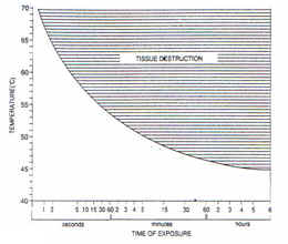
Fig. 2. ПТЕЕПЭ ГыУтНУАЃПЁ ЕћИЅ СЖСїЦФБЋ
ЁЁ
Table 2. ЖпАХПю ЙАМгПЁМ РќУўШЛѓРЛ РЯРИХГМі РжДТ ПТЕЕ
|
Time |
Temperature ( ЁЩ ) |
|
1 second |
70 |
|
2 seconds |
65.6 |
|
10 seconds |
60 |
|
30 seconds |
54.5 |
|
1 minute |
52.7 |
|
10 minute |
48.9 |
(2) ПАСѕЙнРРПЁ РЧЧи УЪЗЁЕЧДТ МеЛѓ(Inflammation mediated injury)
( МіЛѓШФ 1-3РЯ)ШЛѓРИЗЮ ШАМКШЕШ ПАСѕЙнРРРЧ ЕЖМК СЖСЄБт(toxic mediators)ПЁ РЧЧи Дѕ ИЙРК СЖСїМеЛѓРЬ ЙпЛ§ЕШДй. ЙАЗа УЂЛѓФЁРЏРЧ АњСЄПЁ ПАСѕЙнРР(inlammatory response)ДТ ЧЪПфЧЯСіИИ АњЕЕШї Л§МКЕШ ЛъШСІ(oxidants), ДмЙщСњКаЧиШПМв(protease) ЕюРК И№ММЧїАќРЧ ГЛЧЧ(endothelium)РЧ МеЛѓ, ЧЧКЮ ММЦї МеЛѓЕюРЛ ДѕПэ НЩЧЯАд ЧбДй. ДмЙщСњКаЧиШПМв(protease)ДТ ФЁРЏЕЧДТ СЖСї(healing tissue)ИІ МеЛѓНУХАИч, МКРхРЮРк(growth factor)ИІ ЙЋЗТШЧЯИч, ЛъШСІ(oxidants)ДТ ММЦїИІ СзРЬИч, ДмЙщСњРЛ КЏМК, ПАСѕРЛ ДѕПэ ШАМКШЧЯДТ РлПыРЛ ЧбДй.
ЕћЖѓМ РЬЗЏЧб СЖСЄБт(mediators)РЧ РлПыРЛ СЖБтПЁ ЙцСіЧв ЧЪПфАЁ РжДй.(3) ЧуЧїПЁ РЧЧи РЏЕЕЕЧДТ ММЦї МеЛѓ( Ischemia induced cell injury )
МеЛѓЙоРК И№ММЧїАќРК АшМг ЧїРќРЬ СјЧрЕЧИч Дѕ НЩЧб ЧуЧї(ischemia)ПЭ СЖСїБЋЛчИІ РЏЕЕЧбДй. РќНХРћРЮ ЧїОаАЧЯПЭ БЙМвРћРЮ ЧїОзМјШЏ РхЧи ПЊНУ КёНСЧб ШПАњИІ УЪЗЁЧбДй.
(4)Delayed injury
УЪБт ШЛѓ Йз mediatorsРЧ МеЛѓШФПЁЕЕ СіМгЕЧДТ СЖСїРЧ МеЛѓРЬ ПУМі РжДй.
ШЛѓКЮРЧ АЁЧЧ, ММБеС§ЖєЧќМК(bacterial colonization), БтАшРћ ПмЛѓ(mechanical trauma), НЩСіОюДТ ЕЕЦїЧб ПмПыЧзБеСІ(topical antibacterial agent)ЕюПЁ РЧЧиМЕЕ ЙпЛ§ЕШДй.ЖЧЧб ЛѓУГКЮРЇРЧ АњЕЕЧб ШЃСпБИ(neutrophil) Йз БиНЩЧб ДмЙщСњ КаЧиШАМК( proteolytic activity)ЕЕ УЂЛѓФЁРЏИІ СіПЌНУХВДй.
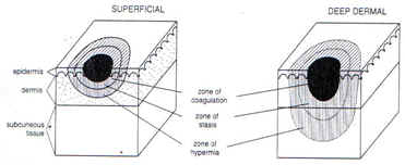
(5)ШЛѓМеЛѓРЧ 3 СіПЊ(Three Zone of Injury)
a.РРАэДы(zone of coagulation or zone of tissue necrosis) КёАЁПЊРћ ММЦїМеЛѓ КЮРЇ
b.СЄУМДы(zone of stasis or Zone of tissue injury) 24~48НУАЃ ГЛПЁ ЦЏКАЧб УГФЁ ОјРЬДТ ММЦї БЋЛчАЁ СјЧрЕЧДТ СіПЊ. СіМгРћРЮ ЧЧКъИА ФЇТј( fibrin deposition),ЧїАќМіУр(vasoconstriction), ЧїРќ(thrombosis)ЕюРИЗЮ ЧуЧї(ischemia)РЛ УЪЗЁЧЯПЉ ММЦїИІ СзАдЧЯДТ КЮРЇ.
c.ПяЧїДы(zone of hyperemia) РћРК ММЦїМеЛѓРЬ ЙпЛ§ЕШ КЮРЇЗЮ АЈПА(invasive infection ) ЖЧДТ НЩЧб ПАСѕРЬ ОјРИИщ МіРЯ ШФ ШИКЙРЬ АЁДЩЧб СіПЊ
УЂЛѓРќШЏ(wound conversion);СжЗЮ СпАЃ Йз НЩКЮ 2ЕЕ ШЛѓ(mid-to deep dermal burns)ПЁМ ШЏКЮРЧ ЧїЗљАЈМв, УЂЛѓФЁРЏБтАЃРЧ СіПЌ,
АњЕЕЧб ПАСѕ Йз НЩЧб АЈПАРЬ РжДТ АцПьПЁ ЙпЛ§ЕШДй.Ся ЛѓБт СЄУМДы(zone of stasis) АЁ РРАэДы(zone of coagulation)ЗЮ КЏШЏЕЧДТ АЭРЬДй.
Fig.3.ШЛѓМеЛѓРЧ 3 СіПЊ(Three Zone of Injury)
ЁЁ